
If you have ever sat in the dental chair and heard, “We might take an X-ray today,” it’s normal to wonder what it shows, whether you really need it, and what it means for your health.
So, what is dental X-ray imaging, and why do dentists use it? In simple terms, dental X-rays are diagnostic images that help your dentist see areas they cannot assess properly with their eyes and mirrors alone. Used appropriately, they support earlier detection, clearer treatment planning, and more confident clinical decisions about your care.
What is dental X-ray imaging?
Dental X-rays are images created using a small amount of ionising radiation to show what is happening beneath the surface. They can reveal:
- Tooth decay between teeth
- Infection around the tooth roots
- Bone levels around teeth linked to gum health
- The position of teeth that are developing or impacted
- Hidden problems beneath existing fillings or crowns
A helpful patient-friendly overview is the dental X-rays information from Guy’s and St Thomas’ NHS Foundation Trust, which explains why X-rays may be recommended and what they help your dentist assess.
Why dental X-rays matter
Many dental problems develop quietly. Small cavities, early gum changes, and tiny cracks may not cause symptoms at first. Dental X-rays can help identify potential problems earlier, when further investigation or monitoring may be appropriate.
X-rays also reduce uncertainty by allowing dentists to base decisions on clinical evidence rather than visual inspection alone. This helps ensure that discussions about care are grounded in what is actually happening below the surface.
This is especially useful during initial assessments, where dentists need a clear baseline before recommending or monitoring any care.
When are dental X-rays needed?
X-rays are not taken automatically at every appointment. In the UK, they should be recommended only when they are clinically justified and likely to influence your care.
Common situations where X-rays may be advised include:
- You are attending a new dentist, and recent X-rays are not available
- Your dentist suspects decay between teeth
- You have toothache, swelling, or sensitivity that needs investigation
- Your gum assessment suggests bone levels may need checking
- You have existing fillings, crowns, or root treatments that require review
- A tooth is missing, delayed, or impacted, particularly in younger patients
- Treatment planning is needed for more complex care
If you are unsure why an X-ray has been suggested, it is reasonable to ask what your dentist is looking for and how the image will inform your care.
The different types of dental X-rays
Dental X-rays come in several formats. The type used depends on the clinical question your dentist needs to answer.
Bitewing X-rays
Bitewing X-rays are commonly used in general dentistry. They show the upper and lower back teeth together and are particularly useful for identifying decay between teeth and checking the margins of existing fillings.
They are often used as part of prevention and monitoring. For a clearer explanation of how decay develops, our guide to understanding cavities outlines the process in straightforward terms.
Periapical X-rays
Periapical X-rays show a full tooth from crown to root, including the surrounding bone. These images are useful when investigating pain, infection, or concerns around the tooth root.
Panoramic X-rays
A panoramic X-ray provides an overview of the entire mouth, including the teeth, jaws, and surrounding structures. It is commonly used when assessing wisdom teeth, missing teeth, or certain jaw-related concerns.
3D scans (CBCT) in selected cases
Cone beam CT (CBCT) scans produce three-dimensional images. These are used only when additional detail is required for diagnosis or planning, such as in some implant assessments, impacted teeth, or complex anatomy.
The British Dental Journal guidance on cone beam CT explains how CBCT imaging is used carefully and selectively in dental care.
What happens when you have a dental X-ray
For most patients, dental X-rays are quick and straightforward.
Step 1: Your dentist explains why the X-ray is being recommended and how it may help.
Step 2: A small sensor or film is positioned in your mouth, often using a holder to keep it steady.
Step 3: You keep still briefly while the image is taken. Exposure time is very short.
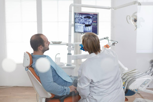
Step 4: Your dentist reviews the image and explains what it shows, often using the screen to guide the discussion.
Most patients find the process quicker and more straightforward than expected. If you feel uncomfortable or anxious, letting your dentist know allows them to make small adjustments where possible.
Are dental X-rays safe?
Dental X-rays involve radiation, so it is sensible to ask about safety. The key point is that X-rays should only be used when they are clinically justified, with exposure kept as low as reasonably practicable.
In the UK, dental X-rays are regulated under the Ionising Radiation (Medical Exposure) Regulations (IR(ME)R). These regulations set legal requirements for justification, optimisation, and professional training.
In practical terms, this means:
- X-rays should only be recommended when there is a clear clinical reason
- The type of X-ray should match the diagnostic need
- Modern digital equipment helps keep exposure levels low
- Your previous imaging history should be considered to avoid unnecessary repetition
If you are pregnant or think you may be, mention this to your dentist. They will weigh the benefits and risks and may delay non-urgent imaging depending on the situation.
Questions patients often ask about dental X-rays
Will X-rays show everything?
No. X-rays are one part of the assessment. They work best alongside a clinical examination, gum checks, and a discussion of your symptoms.
Can I refuse an X-ray?
You can always ask questions and decide what you are comfortable with. If an X-ray is important for diagnosing pain or planning care safely, your dentist will explain why. Choosing not to proceed may limit what can be assessed accurately.
How often should I have X-rays?
There is no universal schedule. Frequency depends on individual risk factors, symptoms, and dental history. Someone with a higher risk of decay may benefit from imaging more often than someone with stable oral health.
A quick comparison of X-ray types
| X-ray type | What it helps assess | When it’s commonly used |
| Bitewing | Decay between back teeth, filling margins | Monitoring and prevention |
| Periapical | Tooth roots and surrounding bone | Pain or suspected infection |
| Panoramic | Whole mouth and jaw overview | Wisdom teeth and planning |
| CBCT | Detailed 3D anatomy | Selected complex assessments |
Planning your care with confidence
Dental X-rays are most useful when they inform a clear and transparent plan. This is particularly important if you are new to the practice or have not had a check-up for some time. The new patient information page explains how initial findings, including X-rays, are reviewed and discussed in a calm, supportive way.
If you would like to ask a question or arrange an appointment, you can contact us via the contact page.
In summary
So, what is dental X-ray imaging for? It is a diagnostic tool that helps dentists assess areas that cannot be seen directly, identify concerns earlier, and plan care more accurately. It should not be used routinely without reason. Instead, it should be recommended when it is clinically appropriate for your individual situation.
The key takeaway is
Dental X-rays support informed decision-making. When used appropriately, they help both you and your dentist base discussions on evidence rather than assumptions.
This article is for general information only and does not replace personalised advice from a dentist who has assessed you in person.